
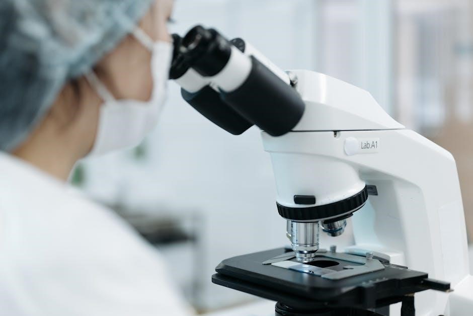
A microscope is an essential tool in science‚ enabling detailed examination of specimens. Its key parts‚ such as the eyepiece‚ objective lenses‚ and stage‚ work together to magnify and focus images for clear observation of cells‚ microorganisms‚ and other microscopic structures.
Overview of Microscope Components
A microscope is composed of several key components‚ each serving a specific function to enhance observation. The primary parts include the eyepiece‚ objective lenses‚ stage‚ and illumination system. The eyepiece‚ or ocular lens‚ magnifies the image formed by the objective lens‚ which focuses light from the specimen. The stage holds the specimen in place using stage clips‚ ensuring stability during observation. The illumination system‚ including the light source and condenser‚ provides and focuses light onto the specimen. Additional components like the diaphragm regulate light intensity‚ while focus knobs adjust the lens position for clarity. Understanding these components is crucial for effective microscope operation and achieving accurate results in scientific investigations.
Importance of Understanding Microscope Parts
Understanding the parts of a microscope is fundamental for its effective use in scientific investigations. Proper knowledge of components like the eyepiece‚ objective lenses‚ and focus knobs ensures accurate specimen observation. Misuse of these parts can lead to poor image quality or damage to the microscope. Recognizing the function of each part‚ such as the role of the condenser in focusing light‚ enhances the ability to achieve optimal magnification and clarity. This knowledge also aids in troubleshooting common issues‚ like blurry images or insufficient illumination; Mastery of microscope components is essential for conducting precise and reliable experiments‚ making it a cornerstone of scientific training and practice.

Major Parts of a Microscope
The major parts of a microscope include the eyepiece‚ objective lenses‚ stage‚ and illumination system. These components work together to magnify‚ focus‚ and illuminate specimens for clear observation.
Eyepiece (Ocular Lens)
The eyepiece‚ or ocular lens‚ is located at the top of the microscope and plays a crucial role in magnifying the image formed by the objective lens. Typically‚ the eyepiece has a magnification power of 10x‚ though higher powers like 15x or 20x are also available. It works in conjunction with the objective lenses to provide the final magnified image of the specimen. In binocular microscopes‚ there are two eyepieces‚ one for each eye‚ ensuring a three-dimensional view. The eyepiece also includes a diopter adjustment to focus the image according to the user’s vision. Proper alignment and focus of the eyepiece are essential for clear and accurate observation of microscopic structures.
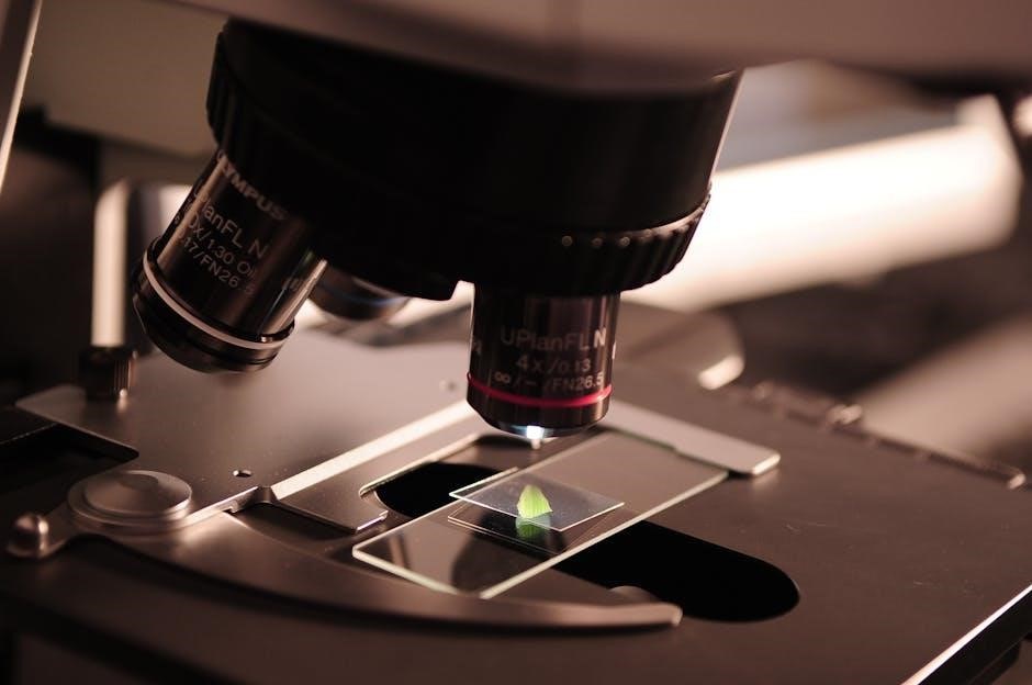
Objective Lenses
Objective lenses are a critical component of a microscope‚ located near the stage and directly above the specimen. They are available in various magnifications‚ such as 4x‚ 10x‚ 40x‚ and 100x‚ with higher magnification lenses providing more detailed views. The objective lens collects light from the specimen and forms an intermediate image‚ which is then magnified by the eyepiece. Each objective lens is designed for specific observational needs‚ with higher power lenses requiring increased light intensity for clarity. Proper alignment and focus of the objective lens are essential for achieving sharp‚ accurate images. The objective lens works in tandem with the eyepiece to produce the total magnification of the microscope‚ enabling users to study specimens at different scales for educational‚ research‚ or diagnostic purposes;
Stage and Stage Clips
The stage is a platform located below the objective lenses where specimens are placed for observation. It is typically made of metal and designed to hold glass slides securely in place using stage clips. The stage clips‚ often spring-loaded‚ ensure the slide remains stable and centered under the objective lens. This stability is crucial for maintaining focus and preventing movement during observation. The stage also allows for precise adjustment of the specimen’s position‚ enabling users to examine different areas of the slide. Its durable construction supports the weight of multiple slides‚ making it a reliable component for both routine and advanced microscopy tasks. Proper use of the stage and clips ensures clear and accurate visualization of specimens under magnification.
Illumination System
The illumination system is a critical component of a microscope‚ responsible for providing light to view specimens. It typically includes a light source‚ such as an LED or halogen bulb‚ located at the base of the microscope. The light travels upward through the condenser lens‚ which focuses the light onto the specimen. The diaphragm‚ part of this system‚ regulates the amount of light that reaches the specimen by adjusting its aperture. Proper alignment of the condenser and light source ensures even illumination‚ which is essential for clear observation. This system is vital for enhancing the visibility and detail of microscopic structures‚ making it easier to study specimens effectively under different magnifications.
Focus Knobs (Coarse and Fine Adjustment)
The focus knobs are essential for adjusting the microscope to bring specimens into clear view. The coarse adjustment knob moves the stage up or down rapidly‚ allowing initial focusing on the specimen. Once the specimen is roughly in focus‚ the fine adjustment knob is used for precise‚ incremental movements to achieve sharp‚ clear images. Proper use of these knobs ensures that the specimen remains centered and well-lit under the objective lens. The coarse knob is typically used at lower magnifications‚ while the fine knob is employed for final adjustments‚ especially at higher magnifications. Together‚ they enable precise control over the focal plane‚ making them indispensable for effective microscopy.
Additional Components of a Microscope
Beyond the primary parts‚ microscopes include a condenser lens‚ diaphragm‚ and light source to manage illumination. Some models feature a binocular or trinocular tube for dual or shared viewing.
Condenser Lens
The condenser lens is a crucial component in a microscope‚ responsible for focusing light onto the specimen. It is typically located beneath the stage and works in conjunction with the diaphragm to regulate light intensity. The condenser lens ensures that the light is evenly distributed across the specimen‚ enhancing image clarity and brightness. Proper adjustment of the condenser is essential for achieving sharp focus and optimal magnification. It is usually aligned with the light source and objective lenses to maximize illumination efficiency. Without the condenser lens‚ the image would appear dim and poorly defined‚ making it difficult to observe details. Regular maintenance‚ such as cleaning‚ is necessary to maintain its effectiveness. This component plays a vital role in the overall functionality of the microscope‚ especially in brightfield microscopy.
Diaphragm
The diaphragm is a critical component located below the stage of a microscope. Its primary function is to regulate the amount of light that passes through the specimen. By adjusting the diaphragm‚ users can control the intensity and focus of the light‚ ensuring optimal illumination for clear observation. It typically consists of an aperture with different-sized holes‚ allowing for varying levels of light transmission. Proper use of the diaphragm enhances image quality by reducing glare and improving contrast. It works in tandem with the condenser lens to focus light effectively. The diaphragm is essential for maintaining image clarity‚ especially when switching between objective lenses with different magnifications. Regular cleaning and maintenance of the diaphragm are necessary to ensure its functionality and the overall performance of the microscope. This component is vital for achieving precise and detailed observations in microscopy.
Light Source
The light source is a fundamental component of a microscope‚ providing the illumination necessary for observing specimens. Typically located at the base of the microscope‚ it emits light through the condenser and onto the sample. Modern microscopes often use LED or halogen bulbs for consistent and adjustable brightness. The light source works in conjunction with the diaphragm and condenser to regulate light intensity and focus. Proper adjustment of the light source ensures clear visibility and optimal contrast‚ enhancing the detail of the specimen. It is essential for achieving sharp‚ well-illuminated images‚ making it a cornerstone of microscope functionality. Regular maintenance‚ such as replacing bulbs or cleaning lenses‚ ensures the light source remains effective. This component is vital for the overall performance of the microscope in various scientific and educational settings.
Binocular or Trinocular Tube
The binocular or trinocular tube is a critical component of a microscope‚ designed to hold the eyepieces in place and align them with the objective lenses. Binocular microscopes have two eyepieces‚ allowing users to view specimens with both eyes‚ while trinocular microscopes include a third port for attaching a camera or other imaging devices. The tube ensures proper spacing between the eyepieces and maintains focus alignment‚ providing a comfortable viewing experience. Trinocular tubes are particularly useful in educational or professional settings‚ enabling image capture or sharing observations. The design of the tube supports ergonomic viewing‚ reducing eye strain during extended use. This versatility makes binocular and trinocular tubes essential for both routine microscopy and advanced applications‚ enhancing functionality and user convenience.

Functions of Microscope Parts
Microscope parts work together to magnify‚ illuminate‚ and focus specimens‚ enabling clear observation and analysis under controlled conditions for scientific study and research purposes.
Magnification Process
The magnification process involves the combined effort of the eyepiece and objective lenses. The objective lens focuses light from the specimen onto the eyepiece‚ which further enlarges the image. Common magnifications include 4x‚ 10x‚ 40x‚ and 100x for objective lenses‚ with eyepieces typically at 10x. The total magnification is calculated by multiplying the two lenses’ powers. For instance‚ a 40x objective lens paired with a 10x eyepiece results in 400x total magnification. Lower magnifications provide a broader view‚ while higher powers reveal finer details. This process is crucial for observing microscopic structures clearly in scientific and educational settings‚ enabling detailed analysis of cells‚ microorganisms‚ and other small specimens.
Specimen Holding and Positioning
The stage and stage clips are essential for securing and positioning specimens under the microscope. The stage provides a flat platform to place slides‚ while the clips hold them firmly in place. Adjustment knobs allow precise movement of the stage‚ enabling accurate positioning of the specimen under the objective lens. Properly placing the specimen on the slide and covering it with a coverslip ensures even illumination and protection. This setup is crucial for clear observation‚ as it keeps the specimen stable and centered‚ allowing users to focus effectively. Without proper specimen holding and positioning‚ achieving sharp and accurate images would be challenging‚ making this process vital for successful microscopy.
Light Management and Focus
Effective light management and focus are critical for clear microscopy. The illumination system‚ including the light source and condenser‚ provides and focuses light onto the specimen. The diaphragm adjusts light intensity‚ optimizing visibility. Coarse and fine adjustment knobs move the stage or objective lenses to bring the specimen into sharp focus. Proper alignment of the condenser ensures even light distribution‚ enhancing image clarity. Adjusting the light and focus correctly prevents eyestrain and allows for detailed observation of specimen structures. These features work together to deliver well-illuminated‚ focused images‚ making microscopy an effective tool for scientific exploration and education.

Using the Microscope Effectively
Using a microscope effectively involves proper preparation‚ focusing‚ and specimen positioning. Start with low magnification‚ then gradually increase for detailed observation‚ ensuring clear and precise results.
Preparing the Microscope for Use
To prepare the microscope‚ place it on a stable surface and ensure the power cord is securely connected. Adjust the diopter setting if using a binocular microscope. Place the prepared slide on the stage‚ securing it with stage clips. Rotate the scanning (4X) objective lens into position and use the coarse adjustment knob to lower the stage. Focus the specimen using the coarse knob until it is in clear view. Ensure the light source is properly aligned and the condenser is adjusted for optimal illumination. Clean the stage and lenses before use to avoid contamination. Proper preparation ensures accurate observations and extends the microscope’s longevity.
Focusing the Specimen
Focusing the specimen is critical for clear observation. Start by rotating the low-power objective lens (4X) into position. Use the coarse adjustment knob to bring the specimen into focus. Once the image is visible‚ switch to the fine adjustment knob for sharper clarity. Ensure the light source is properly aligned‚ and the condenser is adjusted for optimal illumination. If using a binocular microscope‚ adjust the diopter setting for both eyepieces to match your vision. Proper focusing enhances image quality and prevents eye strain. Always focus at low power first to locate the specimen before switching to higher magnification. This step ensures efficient and accurate observation of microscopic structures.
Switching Between Objective Lenses
Switching between objective lenses on a microscope is essential for varying magnification levels. Start by focusing the specimen with the lowest power lens (e.g.‚ 4X) using the coarse adjustment knob. Once the image is clear‚ rotate the nosepiece to select the desired lens (e.g.‚ 10X‚ 40X‚ 100X). After switching‚ use the fine adjustment knob to refine the focus without moving the stage. Avoid touching the slide or stage during the process to prevent damage. Higher magnification lenses provide more detail but reduce the field of view‚ so it’s crucial to locate the area of interest at lower magnification first. Always follow this procedure to ensure clarity and consistency in observations. Proper handling maintains both the specimen and the microscope’s integrity.
Maintenance and Care of Microscope Parts
Regular cleaning of lenses with soft cloths prevents debris buildup. Store the microscope in a dry‚ cool place to avoid damage. Handle with care to ensure longevity and precision.
Cleaning the Lenses
Cleaning microscope lenses is crucial for maintaining optical clarity and performance. Use soft‚ lint-free lens tissue or microfiber cloths to gently wipe away dust and debris. Avoid harsh chemicals or abrasive materials‚ as they can scratch the glass. For stubborn smudges‚ lightly dampen the cloth with distilled water‚ but never apply liquid directly to the lenses. Regular cleaning prevents contamination and ensures sharp‚ clear images. Always clean the eyepiece and objective lenses separately to avoid transferring dirt between them. Store cleaning supplies near the microscope for convenience. Proper lens care extends the lifespan of the microscope and guarantees accurate observations during use.
Storing the Microscope Properly
Proper storage of a microscope is essential to maintain its functionality and longevity. Always cover the microscope with a dust cover or plastic bag when not in use to protect it from dust and moisture. Store it in a cool‚ dry place‚ away from direct sunlight and vibrations. Ensure the microscope is clean and dry before storage to prevent contamination or mold growth. Avoid leaving the microscope assembled‚ as this can strain the mechanical parts; Remove any slides or specimens and store them separately. Secure the microscope in a stable position to prevent accidental movement. For added protection‚ store it in its original case or a padded storage container. Regularly inspect the microscope before and after storage to ensure it remains in optimal condition. Proper storage practices help preserve the microscope’s optical quality and extend its lifespan.
Troubleshooting Common Issues
Common microscope issues often arise from improper use or maintenance. Blurry images may result from incorrect focus‚ dirty lenses‚ or improper slide preparation. If the image is too dark‚ check the light source intensity or ensure the condenser is properly aligned. Low magnification power can be addressed by switching to a higher-power objective lens. Mechanical issues‚ such as stiff focus knobs‚ may indicate over-tightening or lack of lubrication. Dust or debris on lenses can cause distorted images‚ so regular cleaning is essential. If the microscope fails to focus‚ verify that the stage clips are secure and the specimen is centered. For persistent problems‚ refer to the user manual or consult a professional. Troubleshooting these issues ensures optimal performance and extends the microscope’s lifespan.
Advanced Features of Modern Microscopes
Modern microscopes feature digital capabilities‚ specialized objective lenses‚ and automated focus and illumination systems‚ enhancing precision and efficiency in scientific and industrial applications.
Digital Microscopes
Digital microscopes integrate advanced technology‚ combining traditional optics with digital imaging. They feature built-in cameras‚ allowing users to capture high-resolution images and videos of specimens. These microscopes often include LCD screens for direct viewing and software for image analysis‚ enhancing documentation and sharing capabilities. Connectivity options like USB or HDMI enable real-time display on computers or projectors‚ making them ideal for educational settings. Digital microscopes also offer adjustable magnification‚ brightness‚ and focus controls via software‚ streamlining the observation process. Their versatility and modern features make them indispensable tools in education‚ research‚ and industrial applications‚ providing precise and shareable results.
Specialized Objective Lenses
Specialized objective lenses are designed for specific applications‚ offering enhanced imaging capabilities. Phase-contrast lenses improve visibility of transparent specimens‚ while fluorescence lenses highlight specific structures using fluorescent dyes. Polarized light objectives are used for analyzing crystalline materials. These lenses often feature advanced coatings and designs to minimize aberrations and optimize image clarity. They are tailored for disciplines like biology‚ medicine‚ and materials science‚ where precise observations are critical. The choice of objective lens depends on the specimen type‚ desired magnification‚ and required resolution. With their unique properties‚ specialized objectives expand the versatility of microscopes‚ enabling researchers to explore complex samples in greater detail. Their construction involves high-quality glass and precise engineering to deliver superior performance.
Automated Focus and Illumination
Modern microscopes often feature automated focus and illumination systems‚ enhancing efficiency and precision. These systems use motorized stages and focus sensors to automatically adjust the specimen’s position and maintain sharp images. Automated illumination adjusts light intensity and wavelength‚ optimizing visibility for various techniques like fluorescence microscopy. These advancements reduce manual adjustments‚ minimizing human error and saving time. Automated focus is particularly useful for time-lapse imaging and multi-sample analysis. The integration of software-controlled illumination ensures consistent lighting conditions‚ improving image quality and reproducibility. These features are invaluable in research and diagnostics‚ enabling users to focus on analysis rather than instrument setup. Automated systems streamline workflows‚ making microscopy more accessible and efficient for both novices and experts.
A microscope is a vital scientific tool‚ with its parts working harmoniously to reveal microscopic details. Understanding its components enhances its effective use in research and education.
A microscope is composed of essential parts that work together to provide magnified views of specimens. The eyepiece (ocular lens) magnifies the image‚ while objective lenses focus light onto the specimen. The stage holds the slide securely‚ and stage clips keep it in place. The illumination system‚ including the light source‚ condenser‚ and diaphragm‚ manages light for optimal visibility. Focus knobs (coarse and fine adjustments) enable precise specimen alignment. Additional components like the binocular or trinocular tube enhance usability. Understanding these parts is crucial for effective microscope operation‚ allowing users to prepare slides‚ focus accurately‚ and switch between magnifications seamlessly. Proper maintenance ensures longevity and functionality of these critical components.
Final Tips for Effective Microscope Use
To maximize the effectiveness of a microscope‚ start with low magnification to locate the specimen and gradually increase as needed. Properly adjust the focus knobs‚ beginning with coarse adjustments and refining with fine tuning. Ensure the light source is optimized‚ using the condenser and diaphragm to control brightness and contrast. Always handle slides and lenses with care to avoid damage. Regular cleaning of lenses and the stage is essential for maintaining clarity. Store the microscope in a dry‚ cool place to prevent damage from environmental factors. Practice proper techniques for switching objective lenses to avoid disrupting the specimen. By following these guidelines‚ users can achieve precise observations and extend the microscope’s lifespan.